Anatomy of the eye
Anatomy of the eye
The eyes are important organs in the sensory nervous system. Human Eyes are biological cameras in which the light transforms into visual information.
The internal structures of the eye consist of three layers of tissue arranged concentrically: the outer, middle, and inner coat. The sclera and cornea make up the exterior layers. The uvea is the vascular layer in the middle, subdivided into the iris, ciliary body, and choroid. The retina constitutes the innermost layer and is made up of nervous tissue.
The structural components of the eye include:
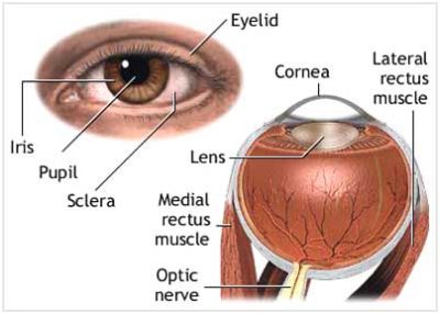
Eyelids: they consist of upper and lower eyelids. They are thin folds of skin that cover the upper and lower parts of the eyeball respectively.
Anterior chamber: it is between the cornea and iris and is filled with a watery fluid called aqueous humour that helps to keep the eyeball inflated.
Posterior chamber: it is between the iris and the lens and is filled with a watery fluid called aqueous humour that helps to keep the eyeball inflated.
Vitreous chamber: it is between the lens and the retina and is filled with a thicker fluid called vitreous humour that helps to keep the eyes inflated.
Vitreous humour. A clear, jelly-like substance that fills the back part of the eye.
Aqueous humour: The clear liquid inside the front part of the eye. It nourishes the eye and keeps it inflated.
Ciliary body: it is behind the iris and connects the choroid to the iris. It helps the eye to focus on different distances. It also provides the aqueous fluid inside the eye.
Conjunctiva: it is a thin mucous membrane that covers the sclera. It protects and lubricates the eye by providing mucus and tears. It prevents microbial entrance into the eye and plays an important role in immune surveillance.
Blood vessels: They are tubes (arteries and veins) that carry blood to and from the eye.
Carucle: A small, red portion of the corner of the eye that contains modified sebaceous and sweat glands.
Sclera: it is the white visible part of the eyeball that protects and covers most of the outside of the eyeball. The muscles attached to the sclera help move your eyeball up and down and side to side.
Choroid: it is between the retina and the sclera. It helps to prevent blurry vision by absorbing the excess light that was entered the eye.
Iris: it is the colored portion of the eye located in front of the lens and behind the cornea. It regulates the amount of light entering the eye. The iris controls the widening and narrowing (dilation and constriction) of the pupil.
Cornea: it is a clear, transparent, and dome-shaped surface in front of the eyeball. It refracts the light entering the eye onto the lens and then transmits it onto the retina.
Lens: it is a transparent structure that is located directly behind the iris. It focuses the entering light rays onto the retina.
Pupil: it is the circular opening in the middle of the iris through which light passes to the lens of the eye. It regulates the amount of light entering the eye by varying its size automatically.
Optic disc: optic disc or optic nerve head is the circular area in the back of the eye where the optic nerve connects to the retina. it is the visible portion of the optic nerve.
Optic nerve: it is located in the back of the eye and transfers all the visual information to the brain.
Retina: it is a light-sensitive layer composed of light-sensitive cells known as cone and red cells. These collections of cells line the inside of the back of your eye. The retina converts light into electrical impulses or neural signals.
Macula: it is a yellow spot located in the center of the retina. it is responsible for detailed central vision and enables us for doing tasks that require central vision such as reading and driving.
Fovea: it is the central portion of the macula and is responsible for central vision.
Rod cells: they are light-sensitive cells in the retina. they function best in dim light.
Cone cells: they are light-sensitive cells in the retina. they function best bright light.